Ultra-Sound contrast imaging is used to visualise perfusion of various tissues in living animals. The aim is to quantitate vascularisation of tumours with or without various radiation or chemical treatments in comparison to normal tissue .
Series of (500 images per scan) are acquired with XXXXX instrument and saved as DICOM image stack.
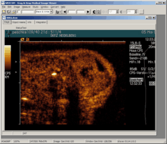
bfDICOM is a generic DICOM viewer allowing
to analyses DICOM image stacks, measure and integrate multiple regions of
interest (ROIs). Multiple single or image-stach DICOM files may be loaded and
analysed simulataneously.
User may define up to 8 freehand regions. These regions are integrated
across the whole image stack. Basic 1-d signal processing allows to
automatically detect inflation / deflation of contrast media (micro-bubbles) and
compute perfusion parameters.
Data may be saved as tab delimited data files and may be
recalled later on. Tab-delimited data files may be easily loaded in to
spreadsheet programs or data bases.
bfDICOM is a monolithic Windows application and does not
require installation nor any supporting libraries or SDKs.
bfDICOM was developed based on ezDICOM medical viewer, copyright
(c) 2002, Wolfgang Krug and Chris Rorden, all rights reserved.
For more details go
here